For many transgender men and nonbinary individuals, phalloplasty is an important and affirming step in aligning their physical body with their gender identity. While traditional approaches to phalloplasty have evolved significantly over time, they often come with visible scars that can carry both physical and emotional weight. That is where Single Scar Phalloplasty (SSP) offers a meaningful advancement.
Developed by Align Surgical’s Dr. Dev Gurjala, SSP is a novel approach designed to minimize scarring while maximizing surgical outcomes, aesthetics and overall well-being.
What Makes SSP Different?
Traditional forms of phalloplasty involve large, visible scars on the forearm, thighs or other parts of the body. SSP, in contrast, confines the visible scarring to a single line on the thigh, without requiring large skin grafts, secondary donor site scars, or a visible incision between the leg and the pubic area in most cases. Additionally, SSP surgery can be performed with or without urethral reconstruction, allowing patients to choose whether to continue urinating while sitting or undergo a urethral reconstruction, which enables them to urinate from a standing position.
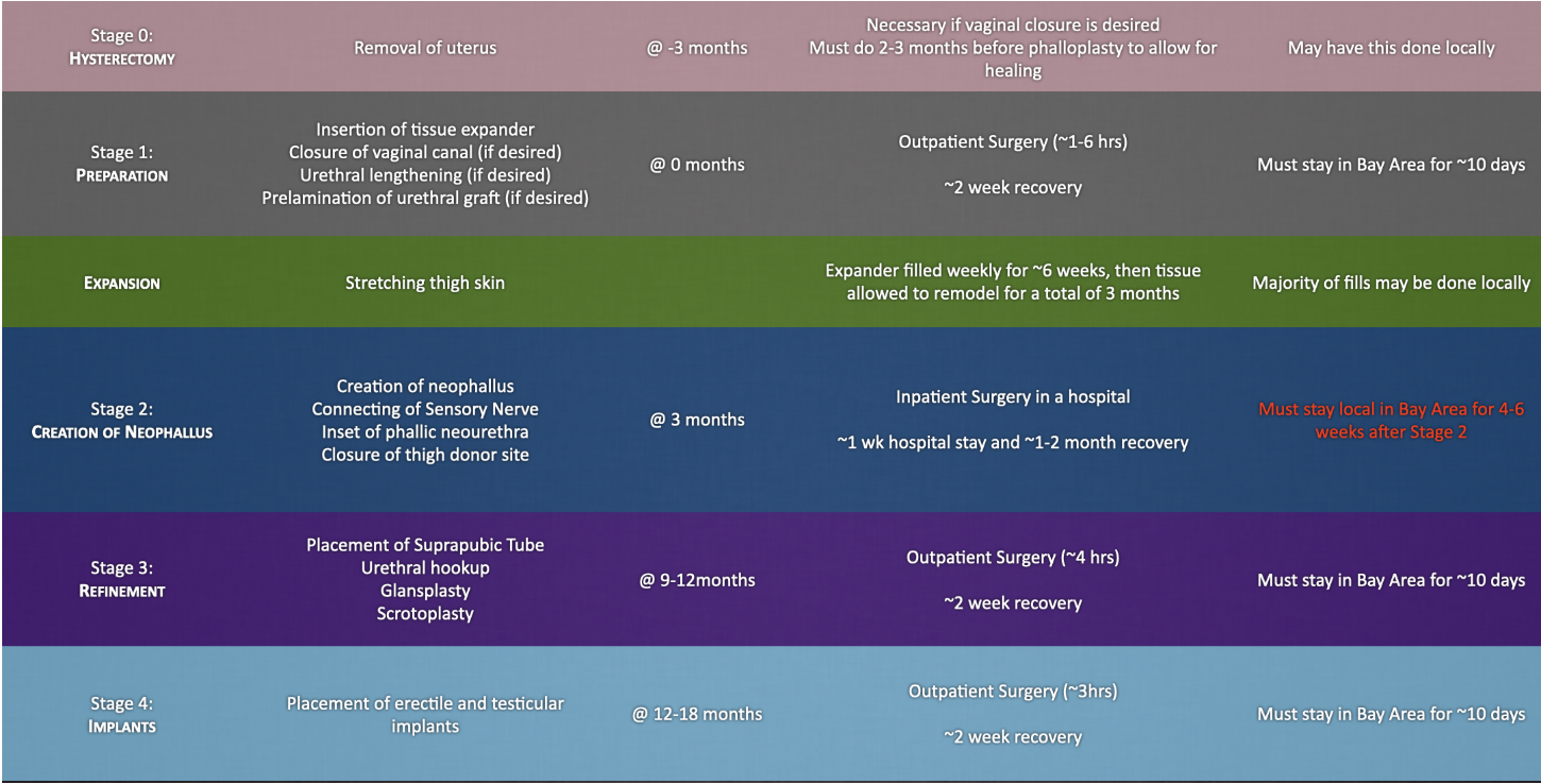
SSP draws from a wide range of reconstructive techniques – such as microsurgery, tissue expansion and prelamination – to construct a functional penis with strengthened blood flow and aesthetic appeal. Aimed at also reducing complications, SSP is performed in stages, allowing for full healing between stages, and personalized pacing that provides flexibility for those with busy schedules.
Phalloplasty Before & After
This gallery contains images intended for adult audiences.
VIEW PHOTOSWhat are the Advantages of SSP?
Minimization of Scars: With SSP, scarring is minimal compared to traditional procedures. With this type of surgery, there is one single line of closure of the thigh donor site, avoiding large skin grafts, and scars connecting the donor site to the pubic area. Scarring from erectile device insertion is limited to a small vertical incision which is usually well-hidden by pubic hair.
Maximizing Tissue Health: The staged approach used in this procedure, such as the delayed tissue expansion or urethral anastomosis stages, allows patients to benefit from increased blood supply and healing while reducing the chances of necrosis (death of skin).
Anatomic Fidelity: With thicker thigh skin, increased connective tissue within thigh fat, and the presence of the tissue expander capsule, patients can enjoy a more natural firmness and texture of the neophallus while minimizing atrophy over time. The best part? The thinner thigh flap also prevents a nonanatomic girth result.
No Need for Preoperative Hair Removal: SSP uses vaginal lining for the neourethra, rather than hair-bearing skin, meaning one less step as part of the pre-operative process.
Contact us to see if this procedure is right for you
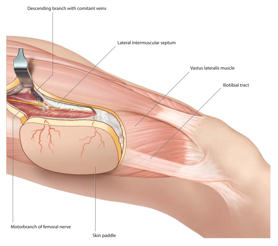
(Illustration 1: SSP Concept)
How SSP Works
SSP begins with a simple evaluation called the pinch test, which involves measuring the thickness of the skin and fat at the mid-thigh. The thickness of this tissue must be about 1 to 1.5 cm to ensure that the eventual flap can be safely tubularized into a neophallus. If the tissue is too thick in this area, it can compromise blood flow to the neophallus or result in a neophallus with an overly large and non-anatomic girth.
Stage 1: Preparation for Phalloplasty
Once cleared, we insert a tissue expander – a device placed under the skin of the thigh through a small (~5 cm) incision. To reduce visible scarring, the incision used to place the expander is later incorporated into the creation of the flap in Stage 2.
Primary risks of Stage 1: vaginal bleeding (rare), leak from urethral lengthening (rare)
Optional Procedures in Stage 1: For those who desire it, this stage may also include removal of the vaginal lining (Vaginectomy), creation of a urethral channel in the thigh using the vaginal lining (Neourethral Prelamination) or relocation of the urethra’s exit point closer to the future neophallus (Urethral Lengthening).
- Vaginectomy: For patients wishing to close the vaginal canal, Stage 1 starts with removing the lining fully from the vaginal canal, followed by closing the vaginal canal (a procedure called Colpocleisis). This offers greater long-term reliability of the vaginal closure, and reduces the risk of dysphoria, infection or chronic buildup that can happen with less comprehensive approaches.
- Note: If a patient wishes to have the vaginal canal closed, hysterectomy 2-3 months before phalloplasty is required
- Neourethral Prelamination: If standing urination is a goal, the vaginal lining removed during vaginectomy can be repurposed to build a neourethra inside the thigh tissue in a process called prelamination. This process gives the future urethra time to heal before being connected later. The vaginal lining can be well-suited for this purpose, as it is elastic, has multiple cell layers and is accustomed to being in a moist environment, unlike external skin.
- Urethral Lengthening: To prepare for future connection between the natal urethra and the neourethra, the urethral exit point is moved forward using a flap from the roof of the vaginal canal. This procedure avoids circular joints, which are prone to narrowing (stricture) and leaks, and instead creates a smooth, straight transition.
Expansion Phase
Over the next three months, the tissue expander is filled weekly with saline through a small port located above the knee. This process slowly stretches and thins the thigh tissue until there is enough surface area to create the neophallus and fully close the thigh donor site as a single line. Tissue expansion has an added benefit of hypervascularizing (increasing blood flow to) the flap, decreasing the chance of necrosis (death of body tissue).
Important Notes:
- Hair removal from the thigh is not done during tissue expansion due to concern for infection, which is the main risk while the tissue expander is in place.
- During this time, it’s important to avoid heavy exercise and wear loose-fitting clothing (like tear-away pants) to accommodate the growing expansion site.
- It is essential that patients not gain weight during the 3-month tissue expansion period, as this can make the flap too thick to safely tubularize during Stage 2. Patients undergoing this procedure must maintain a healthy diet and adjust caloric intake accordingly.
Primary risks of tissue expansion: infection requiring antibiotics, tissue expander malfunction (leak, malposition, or exposure; rare)
Stage 2: Neophallus Creation
Once tissue expansion is complete, the thigh flap is transformed into a neophallus. This is the most technically intensive part of the SSP process, which is performed over two consecutive surgical days for safety and precision. Patients will be required to stay in the hospital ICU for one week and should expect about a one-month recovery period.
The second stage of SSP consists of the following:
Creation of the neophallus
- The procedure begins with raising the flap, i.e., carefully lifting the pre-expanded thigh tissue while preserving critical blood vessels and nerves. This flap remains connected to its original blood supply.
- The flap is then rolled into a cylinder to create the neophallus. This is where the pinch test comes in, as the flap must be thin enough to be safely tubularized without undue tension. If there is too much tension, blood flow can be cut off, increasing the risk for partial necrosis or total flap loss.
- A tunnel is then carefully created under the groin crease, and the flap is passed from the thigh to the pubic area. Tunneling is done to avoid making an incision between the thigh and the pubic area, another effort at reducing scars as much as possible. If tunneling cannot be done safely, an incision is created.
Positioning the neourethra for future connection
- The prelaminated neourethra is then inset (attaching the tissue to its final position) near the previous urethral lengthening. The urethra will be hooked up at a later stage (a “delayed urethral anastomosis”); this is done to minimize the risk of the most common urethral complications: “stricture” (a narrowing in the urethra) and “fistula” (a leak in the urethra).
Coaptation (sewing together) of the sensory nerves
- The sensory nerve brought with the thigh flap is sewn using microsurgery to a nerve that supplies sensation to the clitoris. The goal is to bring tactile (touch) and erogenous (sexual) sensation to the neophallus. It typically takes 1-2 years for nerve sensation to grow into the neophallus, and for us to know if the nerve connection was successful. If nerve growth into the neophallus is successful, patients are anticipated to be able to achieve orgasm from stimulation of the neophallus.
- Importantly, only one of the two main clitoral nerves is used, while the other remains untouched to preserve feeling in the natal clitoris, which is then repositioned and protected at the base of the neophallus. It is expected that orgasm will still be able to be achieved from stimulation of the buried natal clitoris
Closure of the thigh donor site
- Lastly, the thigh wound is closed as a single line scar once more to reduce visible scarring as much as possible. The closure is reinforced at its tightest points with a temporary external skin stretching device called a Dermaclose. On occasion, the skin cannot be fully closed; in this case, the Dermaclose helps bring the skin together over the next 2 weeks, and the closure is completed with a quick return to the operating room, and removal of the Dermaclose device.
- There is still a risk of skin separation after full closure, but this is managed nonoperatively using gauze dressing changes to allow the wound to fully close after 1 – 2 months. In this scenario, the scar is wider, but can be revised to a single line at a later time if desired.
Primary risks of Stage 2: blood flow problems leading to venous congestion, partial necrosis (rare) or total flap loss (rare); failure of sensation to reach neophallus (rare); inability to fully close thigh donor site; wound healing problems at thigh closure site (separation of incision)
Stage 3: Refinement
Stage 3 is focused on refining the anatomy and making key connections for functionality. Typically scheduled about 6 months after Stage 2 (or whenever you’re ready), this stage allows time for healing and tissue stabilization.
Suprapubic tube placement: A suprapubic catheter tube to drain the bladder is placed in case any urinary complications arise during healing.
Urethral hookup (anastomosis): The prelaminated urethra is connected to the lengthened natal urethra using a straight-line closure designed to reduce leaks (fistula) and narrowing (stricture).
Scrotoplasty: Flaps are shaped into a scrotum, which also provides an extra layer of coverage over the new urethral connection.
Glansplasty: A groove is created (the “coronal sulcus”) to separate the head from the shaft of the neophallus, often using a small skin graft from the thigh.
Slit meatoplasty: A slit may be created at the tip to mimic the appearance of a urethral opening, especially for those not undergoing urethral reconstruction.
Primary risks of Stage 3: urethral stricture or fistula, partial necrosis of scrotum (rare), effacement of glansplasty (smoothing out of groove over time)
Stage 4: Implant Placement
Once the neophallus has healed and the sensation has returned (typically 1 – 2 years after Stage 2), you may choose to move forward with implanting an erectile device and testicular implant.
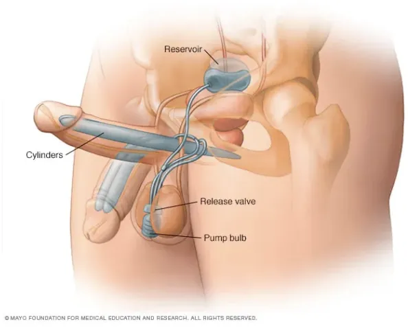
Erectile device insertion: An inflatable penile prosthetic is placed through a small, vertical incision above the neophallus and anchored to the pubic bone. A fluid reservoir is placed in the abdomen, and a pump is inserted into the neoscrotum. The pump serves as one of the testicles.
Testicular implants: If receiving an erectile device, a single testicular implant is placed in the other side of the neoscrotum. For patients not wishing to have an erectile device, two testicular implants may be placed into the neoscrotum.
Primary risks of Stage 4: erectile device malfunction (leak, mechanical failure, or exposure over time), implant malposition, implant infection
(Illustration 2: Erectile Device and Testicular Implant Placement)
Is SSP Right for You?
While SSP offers many advantages, it is not the ideal path for everyone. There are several factors to consider with SSP, including tissue thickness or lifestyle factors that make tissue expansion challenging. And SSP requires patience – typically four stages spanning 1-2 years or more.
At Align Surgical, we believe that with any surgical procedure, patients must have access to all the information they need to make an informed decision and be comfortable emotionally, socially, and logistically. Whether SSP ends up being the best option for you or not, we will help you explore the approach that will achieve your embodiment goals.